Feto-placental angiogenesis is tightly regulated by oxygen tension, circulating factors and, in a paracrine manner, by neighbouring cells.
A rich source of angiogenesis regulating factors are placental trophoblasts that form the outer surface of the placenta and border the placental stroma, and placental tissue macrophages, the Hofbauer cells, that are often located in close proximity to the endothelium. In order to identify the paracrine interplay between feto-placental endothelial cells with trophoblasts and Hofbauer cells, we isolated these cells to investigate the effect of conditioned medium on feto-placental angiogenesis. Thus, we identified the trophoblast derived molecule PEDF as a negative regulator of angiogenesis that limits vascular growth at the end of pregnancy. Moreover, we demonstrated that Hofbauer cells – similar to tissue macrophages in adult organs - regulate feto-placental endothelial function in a paracrine manner. Maternal factors may alter these regulatory circuits and modify feto-placental angiogenesis.
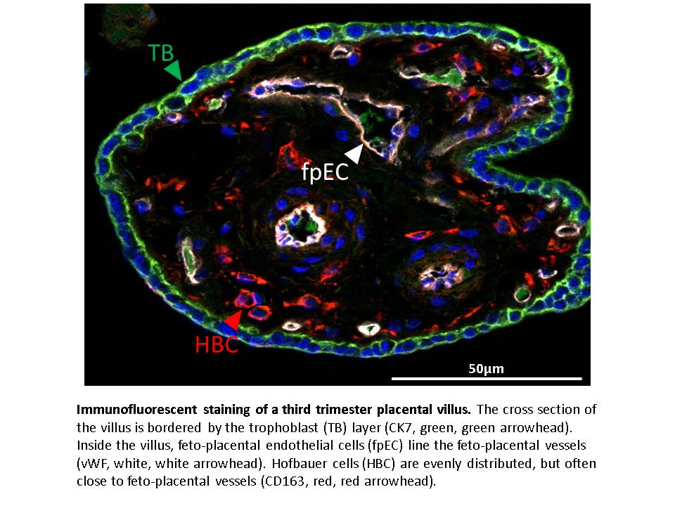
Immunofluorescent staining of a third trimester placental villus.
The cross section of the villus is bordered by the trophoblast (TB) layer (CK7, green, green arrowhead). Inside the villus, feto-placental endothelial cells (fpEC) line the feto-placental vessels (vWF, white, white arrowhead). Hofbauer cells (HBC) are evenly distributed, but often close to feto-placental vessels (CD163, red, red arrowhead).
Literatur:
Loegl et al, Angiogenesis 2016
Loegl et al., Reproduction 2016
Lassance et al., Histochen Cell Biol
Ghaffari-Tabrizi et al., Cells Tissues Organs 2015