Ex vivo perfusion of the placenta
 Birgit Hirschmugl
 Christian Wadsack
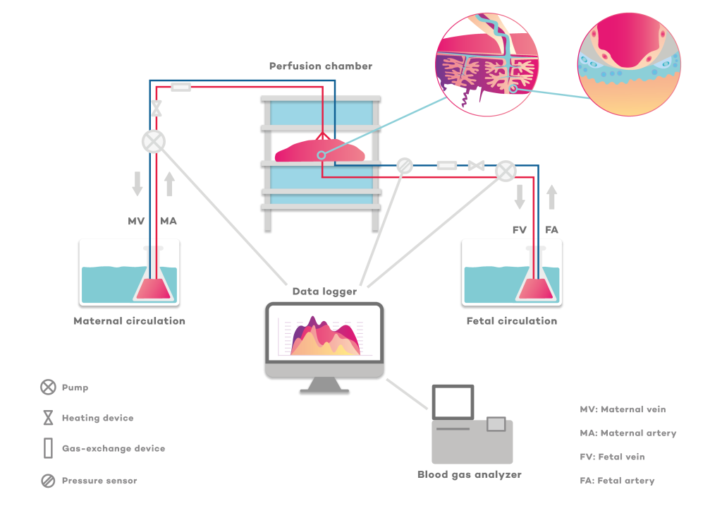
Ex vivo perfusion of the placenta was developed around 40 years ago. It is a delicate method that requires well-trained, experienced personnel. A small functional unit of the placenta, called a cotyledon, is removed from the placenta. A pair of cannulated fetal blood vessels, artery and vein, and a buffer reservoir containing a cell culture medium that keeps the placental cells metabolically functional are then used to mimic the fetal circulation. Since the placenta is only loosely perfused with maternal blood, the maternal circulation is mimicked by inserting blunt cannulas through the basal plate of the placenta into the intervillous space. This maternal circulation is also provided by a buffer reservoir.
Depending on the research interest, various nutrients/pharmacological agents etc. can be added to the maternal reservoir and the transfer of such substances to the fetal side (but also back into the maternal reservoir) can be investigated. In recent decades, a large number of perfusion studies have expanded our knowledge of how antibodies, growth factors, hormones and environmental pollutants pass through the placenta. It is also possible to study transport in different maternal metabolic environments by using the placenta from pregnancies affected by pre-eclampsia, growth restriction or diabetes and comparing whether their transport rates are altered compared to uncomplicated pregnancies.